From The Lancet:
"A haemodialysis patient with hypercholesterolaemia was admitted with an ulcer on his left foot. Shortly after admission he complained of generalised aching and difficulty in mobilising.
Osteomyelitis complicating his foot ulcer was suspected and a Tc 99 -labelled diphosphonate bone scan was done. The scan showed uptake into damaged muscle"
Click here to see the images from The Lancet website.
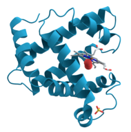
Model of helical domains in myoglobin, the protein linked to kidney damage in rhabdomyolysis. Image source: Wikipedia, public domain.
Rhabdomyolysis is the rapid breakdown (lysis) of skeletal muscle tissue (rhabdomyo) due to injury to muscle tissue. Rhabdomyolysis and its complications are major problems in people who are injured in disasters such as earthquakes and bombing. The disease and its mechanisms were first elucidated in the Blitz of London in 1941.
References:
Visualising rhabdomyolysis. Stephen Walsh et al. The Lancet, Volume 373, Issue 9658, Page 154, 10 January 2009.
Rhabdomyolysis, from Wikipedia, the free encyclopedia.
Related:
Rhabdomyolysis Review, Hospital Physician, 2009 (PDF).
Updated: 05/14/2009
"A haemodialysis patient with hypercholesterolaemia was admitted with an ulcer on his left foot. Shortly after admission he complained of generalised aching and difficulty in mobilising.
Osteomyelitis complicating his foot ulcer was suspected and a Tc 99 -labelled diphosphonate bone scan was done. The scan showed uptake into damaged muscle"
Click here to see the images from The Lancet website.
Model of helical domains in myoglobin, the protein linked to kidney damage in rhabdomyolysis. Image source: Wikipedia, public domain.
Rhabdomyolysis is the rapid breakdown (lysis) of skeletal muscle tissue (rhabdomyo) due to injury to muscle tissue. Rhabdomyolysis and its complications are major problems in people who are injured in disasters such as earthquakes and bombing. The disease and its mechanisms were first elucidated in the Blitz of London in 1941.
References:
Visualising rhabdomyolysis. Stephen Walsh et al. The Lancet, Volume 373, Issue 9658, Page 154, 10 January 2009.
Rhabdomyolysis, from Wikipedia, the free encyclopedia.
Related:
Rhabdomyolysis Review, Hospital Physician, 2009 (PDF).
Updated: 05/14/2009